Hematoxylin and Eosin (H&E) staining is one of the most common techniques used in histology to visualize tissue structures. This method allows researchers and pathologists to examine tissue samples under a microscope. Let's explore how H&E staining works and its significance in medical and scientific studies.
Hematoxylin and Eosin Staining and How Does It Work?
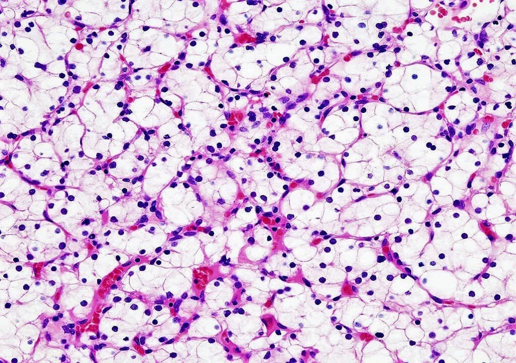
To answer, what is H&E? Hematoxylin and Eosin staining is a two-step process used to color tissue sections. During this staining procedure, the first step involves using hematoxylin, a dye that stains the cell nuclei in either dark purple or blue. The second step uses eosin, which stains the cytoplasm, collagen, and extracellular components in various shades of pink and red. This combination allows for clear visualization of the tissue architecture, enabling the differentiation of cellular components.
This method is essential in both diagnostic and research labs. H&E staining helps distinguish between different tissue types and cellular structures, such as the nucleus and cytoplasm. It is particularly valuable in the detection and classification of diseases, including cancer, where tissue abnormalities need to be studied closely.
Applications of H&E Staining in Medical Diagnosis
Hematoxylin and Eosin staining plays a crucial role in medical diagnostics, especially in pathology. It allows pathologists to analyze tissue samples and diagnose various conditions, including cancer, infections, and inflammatory diseases. The clear contrast between the stained structures makes it easier to identify abnormal cells or tissues.
This method is particularly beneficial for detecting malignancies, as cancer cells often show significant differences in shape, size, and structure when compared to healthy cells. By examining H&E stained slides, medical professionals can assess the tissue's condition, which is vital for developing accurate treatment plans. It also aids in determining the stage of the disease, helping to guide medical decision-making.
Key Benefits of H&E Staining
H&E staining is preferred in many laboratories for several reasons. The widespread use of this method has made it a cornerstone in tissue analysis, particularly in histopathology. Its ability to differentiate between cell types and reveal structural details makes it an indispensable tool in medical diagnostics.
Advantages of H&E Staining:
- Low-cost and widely available
- High contrast for clear tissue differentiation
- Suitable for a variety of tissues and diseases
- Easy to interpret and analyze
Reputable Biospecimen Provider Ensure Quality
When using H&E staining for tissue analysis, it’s crucial to work with a reputable biospecimen provider. A reliable provider ensures that the tissue samples are of the highest quality and prepared properly for staining. This guarantees that the results from H&E staining are accurate and reproducible, which is vital for both research and diagnosis.
Working with a trusted biospecimen provider also ensures that the tissue specimens are handled according to strict quality standards, minimizing the risk of contamination or degradation. This is especially important in sensitive studies or medical diagnoses, where precision is essential. By partnering with a reputable provider, laboratories can improve the overall quality and reliability of their tissue analysis.
Understanding Hematoxylin and Eosin staining is essential for anyone involved in tissue analysis. Whether the question is, what is H&E? or you are looking to implement the technique, it is a powerful and accessible method for visualizing tissue structures. By utilizing reputable biospecimen providers and ensuring proper techniques, labs can maximize the effectiveness of H&E staining for both diagnostic and research purposes.